
“Dense doesn’t mean doomed. It means your screening plan should be smarter.”
– Wellness Kraft
Introduction
Few medical phrases create instant confusion like: “You have dense breasts.”
Because it sounds like a diagnosis, it sounds like a warning. It sounds like you’re being told something is wrong, without being told what to do next.
Most women immediately ask the same three questions:
- Is this dangerous?
- Does it mean I’m more likely to get breast cancer?
- Do I need extra tests like an ultrasound or an MRI?
And then the internet does what it does best: throws 400 opinions at you, ranging from “Ignore it” to “You must get an MRI every year, or you’re risking your life.”
So let’s slow it down and make it clear.
Breast density is not something you can feel. It’s not about breast size or firmness. It’s something radiologists see on a mammogram, based on how much fibrous and glandular tissue you have compared to fatty tissue.
Why it matters:
- Dense tissue can make it harder for mammograms to spot cancer because both dense tissue and many tumors appear white on the image.
- Dense tissue is also linked to a somewhat higher risk of breast cancer compared to less dense tissue.
But here’s the most important part:
Dense breasts alone do not automatically mean you need an MRI or additional imaging. Many women with dense breasts are still appropriately screened with mammography, especially when combined with a good risk assessment and, sometimes, 3D mammography.
This article will help you determine what’s reasonable, what’s overkill, and which questions to ask so you leave your next appointment feeling informed rather than confused.
What “Dense Breasts” Actually Means
On mammogram reports, breast density is usually described in four categories:
- Mostly fatty
- Scattered areas of fibroglandular density
- Heterogeneously dense
- Extremely dense
The last two categories are what most people mean when they say “dense breasts.”
Dense breasts are common. You’re not an outlier. Many women, especially younger women and some premenopausal women, have dense tissue. Density can also change with age, hormonal changes, pregnancy, and menopause.
Two key facts to keep in your head at the same time
- Dense tissue can hide cancers on a mammogram.
- Dense tissue is also associated with a higher risk of breast cancer compared to non-dense tissue.
That’s it. That’s the core.
The following steps are about reducing the chance of “missed cancers” while also avoiding unnecessary anxiety, false alarms, and extra procedures that don’t benefit you.
Megan’s “Do I Need an MRI Now?” Moment
Megan is 42 and lives in Seattle. She’s healthy, busy, and the kind of person who schedules medical appointments like she schedules oil changes: responsibly, but without drama.
She gets her first screening mammogram and later sees a message in her portal:
“Your breasts are heterogeneously dense.”
That’s all her brain catches. Dense.
Megan spirals. She imagines “dense” equals “high risk.” She starts googling. She reads a thread where someone says mammograms are “useless” for dense breasts. She sees a video claiming every woman with dense breasts needs an MRI.
By day two, Megan is terrified. Not because she has symptoms. Not because her mammogram found cancer. But because she feels like she just learned there’s a blind spot in her body.
When she finally talks to her clinician, the conversation changes everything:
- Megan has no strong family history.
- No prior chest radiation.
- No known genetic risk.
- No prior breast biopsies with high-risk findings.
- Her mammogram was regular.
Her clinician explains: “Dense breasts matter, but they’re just one part of your risk picture. Let’s calculate your overall risk and then decide if you need anything beyond mammography.”
They do a risk assessment. Megan is classified as average risk. The plan becomes:
- Keep screening.
- Consider 3D mammography (tomosynthesis) if available.
- Reassess risk periodically.
- No automatic MRI, because the chance of false positives and unnecessary biopsies would likely outweigh the benefits for her risk level.
Megan leaves calmer, not because she ignored density, but because she stopped treating it as a verdict and started treating it as information.
That’s the goal.
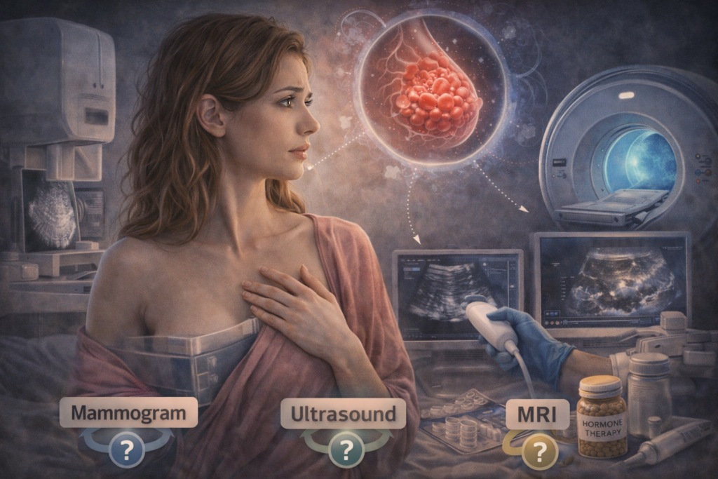
Mammograms, Ultrasound, MRI: What Each One Does Well
Mammogram
This is the standard screening test. It’s the foundation for most women.
Best for:
- Detecting many early cancers and microcalcifications
- Population screening (catching cancers before symptoms)
Limitations:
- Dense tissue can reduce visibility
- Can lead to false positives (callbacks) and anxiety
3D Mammogram (Tomosynthesis)
This is often referred to as “3D mammography.” It captures images in slices, which can reduce tissue overlap.
Why people like it:
- Can improve cancer detection in some women
- Can reduce recall rates in specific settings
- Often feels like a “mammogram, but better,” especially for dense breasts
Ultrasound (Supplemental)
Ultrasound can detect cancers that mammograms might miss in dense tissue, but it also detects many non-cancer findings.
Pros:
- Can detect some additional cancers in dense breasts
- No radiation
Cons:
- Higher false positive rate
- More callbacks and biopsies that turn out benign
- Results can depend on technique and technology
MRI (Breast MRI or Abbreviated MRI)
MRI is very sensitive. It can detect cancers that mammograms and ultrasounds miss, especially in higher-risk women.
Pros:
- Best at finding additional cancers, especially in high-risk women
- It can be life-saving in the right population
Cons:
- More false positives than mammography
- More follow-up testing and sometimes biopsies
- Cost/availability and sometimes contrast considerations
Contrast-Enhanced Mammography (CEM)
This is a newer-ish tool that combines mammography with contrast enhancement. It’s not everywhere, but it’s part of the evolving landscape.
The Most Important Step Most People Skip: Risk Assessment
Breast density is a factor, but it’s not the whole story.
Your real decision is based on your overall risk, including things like:
- family history (especially first-degree relatives)
- known genetic mutations (BRCA1/2 and others)
- Prior chest radiation at a young age
- certain previous biopsy findings
- personal history of breast cancer
- age, reproductive history, and other factors
Here’s the practical truth:
A woman with dense breasts but otherwise average risk may not benefit from MRI the way a woman with dense breasts and high-risk factors does.
So the best approach is:
- Know your density category
- Know your baseline screening schedule
- Calculate risk
- Then decide on “add-ons” like ultrasound or MRI
How to Choose the Right Screening Plan
This is a practical guide, not a rigid rule.
If you are average risk, and your mammogram is normal
- Mammography remains your primary screening tool.
- If available, consider 3D mammography.
- Talk to your clinician about whether a supplemental ultrasound is worth it for you. For some average-risk women, it may add detection. For others, it mainly adds anxiety and extra testing.
If you have dense breasts AND a higher risk (or possibly higher risk)
You should strongly consider a more personalized plan. This is where MRI often becomes more relevant.
Higher risk can include:
- strong family history
- known genetic mutation
- Prior chest radiation
- certain high-risk biopsy findings
- calculated lifetime risk in a higher-risk range
In this category, MRI may be recommended regardless of breast density, depending on your exact risk profile.
If you have symptoms
Whether breasts are dense or not, symptoms change the conversation.
Symptoms that deserve evaluation:
- new lump
- persistent focal pain (one area, consistent)
- nipple discharge (especially bloody or spontaneous)
- skin changes, new nipple inversion
- significant change in size/shape
Screening tests are for people without symptoms. Symptoms often require diagnostic imaging and clinical evaluation.
What to Ask Your Doctor
If you want to walk out with a plan that actually makes sense, ask these:
- What is my breast density category (heterogeneously dense or extremely dense)?
- Based on my history, am I average, intermediate, or high risk?
- Should we do a formal risk assessment?
- Would 3D mammography be better for me?
- Do the benefits of supplemental ultrasound outweigh the downsides in my situation?
- Do I meet the criteria for an MRI recommendation?
- If I conduct additional screening, how often, and which test?
If your clinician can’t answer, consider asking for a referral to a breast specialist or a high-risk clinic for a one-time risk consultation.
The Emotional Side Nobody Talks About
The worst part of dense breast notifications is the psychological effect: now you’re aware there might be a limitation in what the test can see. That creates a background fear even when everything is normal.
So here’s the grounding statement:
Screening is risk management, not risk elimination.
No screening strategy is perfect. The goal is to choose the plan that gives you the best chance of early detection with the least unnecessary harm.
And you’re allowed to choose calm, not constant fear.
Research Insight
Breast density is reported on mammography because dense tissue can reduce mammographic sensitivity and is associated with increased breast cancer risk compared with less dense tissue. The U.S. FDA’s updated MQSA requirements include notifying patients about breast density in mammography reports, and facilities were required to comply beginning September 10, 2024. U.S. Food and Drug Administration+2U.S. Food and Drug Administration+2
Screening recommendations vary by organization, but a major update in the U.S. is the USPSTF 2024 final recommendation for biennial screening mammography for women ages 40 to 74 (Grade B). The USPSTF also states evidence is insufficient to assess benefits/harms of supplemental screening (like ultrasound or MRI) for women with dense breasts after a negative mammogram, which is why risk-based personalization matters. USPSTF+2USPSTF+2
Professional society guidance and emerging evidence indicate that supplemental imaging (ultrasound, MRI, abbreviated MRI, contrast-enhanced mammography) may be useful in certain risk categories, particularly for women at higher risk, while also acknowledging trade-offs such as false positives and downstream testing. ScienceDirect+3ACR Acsearch+3JACR+3
Links (paste as raw URLs in WordPress for auto-clickable sources):
https://www.uspreventiveservicestaskforce.org/uspstf/recommendation/breast-cancer-screening
https://jamanetwork.com/journals/jama/fullarticle/2818283
https://www.fda.gov/radiation-emitting-products/mammography-quality-standards-act-mqsa-and-mqsa-program/important-information-final-rule-amend-mammography-quality-standards-act-mqsa
https://www.fda.gov/news-events/press-announcements/fda-updates-mammography-regulations-require-reporting-breast-density-information-and-enhance
https://www.cancer.org/cancer/types/breast-cancer/screening-tests-and-early-detection/american-cancer-society-recommendations-for-the-early-detection-of-breast-cancer.html
https://acsearch.acr.org/docs/3158166/Narrative/
https://www.jacr.org/article/S1546-1440%2825%2900129-2/abstract
https://pubs.rsna.org/doi/abs/10.1148/radiol.221785
Key Takeaways
- Dense breasts are common and are determined by mammogram appearance, not by feel or size.
- Dense tissue can make cancers more complicated to see on mammograms and is linked to a higher breast cancer risk compared with non-dense tissue.
- Mammography remains the foundation of screening for most women; 3D mammography can be helpful, especially for women with dense breast tissue.
- Ultrasound and MRI can find additional cancers in some women, but they can also increase false positives and unnecessary procedures.
- The smartest next step is risk assessment: dense breasts plus higher risk may justify MRI, while dense breasts alone may not.
FAQs
1) If I have dense breasts, does that mean I will get breast cancer?
No. Dense breasts are a risk factor, not a prediction. Many women with dense breasts never develop breast cancer, and many women with non-dense breasts do. Density is one piece of the risk puzzle. Your age, family history, genetics, prior biopsies, and other health factors matter too.
2) Should every woman with dense breasts get an ultrasound?
Not necessarily. Supplemental ultrasound can find additional cancers, but it can also lead to more false positives, more callbacks, and more biopsies that turn out to be benign. The decision should be based on your overall risk and how you personally weigh the tradeoff between “finding more” and “going through more unnecessary testing.”
3) Is MRI better than ultrasound for dense breasts?
MRI is generally more sensitive than ultrasound, but it’s not automatically the right choice for everyone. MRI is often recommended for women at higher risk (based on genetics, strong family history, certain medical history, or a high calculated lifetime risk). For average-risk women, MRI may lead to more false positives without a clear overall benefit for everyone.
4) Can breast density change over time?
Yes. Density often decreases with age, especially after menopause, but it’s not universal. Hormonal factors, medications, and individual biology can influence it. That’s why your density category may change across different mammograms.
5) What should I do right now if my report says “dense breasts”?
Do three things:
- Ask which density category you have.
- Ask for a basic risk assessment based on your history.
- Discuss whether 3D mammography, ultrasound, or MRI makes sense for your risk level and your peace of mind. The goal is a plan you can stick to without living in panic.
Concluding Thoughts
Dense breasts are not a diagnosis. They’re a data point.
The real win is not chasing every test out of fear. The real win is building a screening plan that fits your actual risk, uses the right tools, and keeps you coming back consistently.
If your report says “dense,” don’t treat it like a cliff. Treat it like a signpost:
Pause, assess your risk, and choose the next step with clarity.


Join the conversation